
 Facebook
  Twitter
  RSS
RSS
Heterochromia
What is Heterochromia?
The word heterochromia is a combination of two Greek words, which mean “different colors”. In the medical field, this term is sometimes used in connection with a skin or hair condition. However, most of the time heterochromia refers to a change in color of a part of the eye, called the iris. This condition is also know as “heterochromia iridum”.
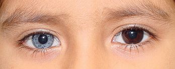
Two different eye colors is a trait in Heterochromia.
Heterochromia dates back to ancient times. Aristotle described the condition initially and he referred to it as “heteroglaucos”. The eastern Emperor Anastasios was called Dicorus, meaning “two-pupiled”, because he had different colored eyes. The difference in color can be caused by distribution in asymmetric concentrations in the iris or due to varying amounts of melanin in the iris tissues.
The development of eye color is established by the time a child is 2 years old, meaning that the iris has become fully pigmented. There are 4 factors that determine eye color:
- Heredity/genetics
- Sympathetic stimulation of the iris melanocytes (cells that produce melanin in the eye),
- Melanocyte stimulating hormone (MSH), and
- Biochemical factors that impact the metabolism of melanin.
An alteration in any of these 4 factors can cause the color of the iris to change. Alterations can occur during fetal development (congenital heterochromia) and/or due to injury or diseases/conditions (acquired heterochromia) that impact any of the 4 factors. In most cases, the color change occurs bilaterally (it impacts both eyes). However, in rare cases, the change in color impacts only one eye. This is called heterochromia.
Types of Heterochromia
What are the types of heterochromia? Heterochromia can occur as either hypo-pigmentation (lack of pigmentation resulting in a lighter colored iris) or hyper-pigmentation (excess pigmentation resulting in a darker colored iris). The difference in the color of the iris is due to too much or too little pigmentation. It can occur in three degrees:
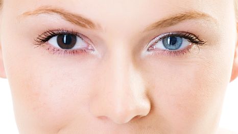
1. Complete Heterochromia
In complete heterochromia the iris of each eye is a completely different color. As you can see in the image below, one eye is light blue and the other is brown.
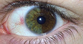
2. Sectoral/Partial Heterochromia
For it to be a sectoral/partial heterochromia, a segment or portion of contrasting colors in the iris must be present. In the image you can see spots of a light brown color in the iris.
Patches of brown color in the eye.
Half the eye in different color.
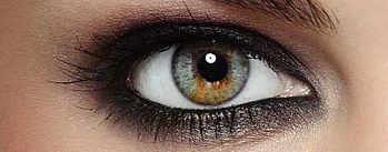
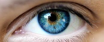
3. Central Heterochromia
The characteristics in central heterochromia is that one color radiates from the pupil (the dark center of the eye), and then shifts to another color. You can see in the image that the pupil is surrounded by a yellow/brown color. This color radiates and shifts to a blue color
Incidence of Heterochromia
How rare is heterochromia? Interestingly enough, heterochromia commonly occurs in dogs! However in humans, it occurs very rarely and is officially classified as a “rare disease” by the Office of Rare Diseases at the National Institute of Health. It is difficult to report an accurate incidence rate since in many cases, the difference of color between the two eyes is so subtle and therefore it goes unnoticed. The NIH estimates that heterochromia iridis affects less than 200,000 people in the US. The estimated incidence of congenital heterochromia is approximately 6 in 1,000 live births.
Causes of Heterochromia
What causes heterochromia? In most cases, heterochromia is hereditary. It is an autosomal dominant trait, meaning that it is likely one of the parents of an affected individual also has heterochromia. Several family members will also have heterochromia. People with hereditary (or congenital) heterochromia are born with different colored iris, called congenital heterochromia. Heterochromia can also be acquired later in life. This can be through an illness, injury, use of certain medications, or aging. For example, areas of iris hypopigmentation are commonly seen in older people; and this may be due to senile iris atrophy simply as part of the aging process. Heterochromia can occur by itself with no other disorders or problems, or it may occur as part of a syndrome or disease. It usually does not impact vision.
Congenital heterochromia has been associated with the following conditions:
- Tuberous sclerosis / Neurofibromatosis: This disease is characterized by development of several benign tumors (such as hamartomas) in multiple organs, including brain, heart, skin, eyes, kidney, lungs, and liver. Affected individuals have a lighter colored iris.
- Waardenburg syndrome: This is an autosomal dominant (hereditary) condition. There are four types of Waardenburg syndrome. Some of the common symptoms include: hearing loss, difficulty straightening joints, wide-set eyes, white patch of hair or early graying of hair, and changes in the pigmentation of the hair, eyes, and skin (many patients with Waardenburg syndrome have very pale blue eyes or different colored eyes).
- Horner’s syndrome secondary to birth trauma or intrauterine trauma. Horner’s syndrome consists of ipsilateral eye lid dropping (ptosis), miosis, and facial anhidrosis. It also can result in hypo-pigmentation of the iris. It is a result of injury to the nerves in the sympathetic chain which innervate the eye. An injury may occur during birth, or the condition can occur later in life due to trauma or disease to the sympathetic nerve. The eye on the affected side has a smaller pupil, ptosis (drooping eyelid), and lighter colored iris.
- Sturge-Weber Syndrome: This syndrome is characterized by a port wine stain and angioma involving the brain and eye. Affected individuals will sometimes have a darker pigmented iris on the same side of their facial angioma. The hyper-pigmentation is a result of a melanin based hamartoma on the surface of the iris.
- Hirschsprung disease: this is a disorder of the gastrointestinal system. It is sometimes associated with sectoral heterochromia, lighter (less pigmented) iris. The hypo-pigmentation is a result of reduced numbers of pigment-producing cells in the affected eye. When heterochromia is present along with Hirschsprung disease, it is likely due to neural crest defects that occurred during fetal development.
- Parry-Romberg syndrome: This syndrome is characterized by hemi-facial atrophy of the skin and tissue around the forehead. Heterochromia is present in some of those affected.
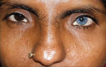
Indian women with Waardenburg Syndrome showing complete heterochromia iridis.
Acquired heterochromia (acquired later in life, after birth) has been associated with the following syndromes or diseases:
- Trauma/injury can cause the iris to atrophy or impact the function of melanocytes, and consequently result in hypo-pigmentation (iris becomes lighter). Bleeding can also impact the color of the iris.
- Acquired Horner’s Syndrome: Deficiency of sympathetic activity interferes with melanin pigmentation of the melanocytes in the stroma of the iris. This also results in the iris becoming lighter colored.
- Herpes simplex: Inflammation of the iritis may result in decreased pigmentation and loss of color of the iris.
- Iron deposition into the iris (due to siderosis or hemosiderosis) may result in darkening of the iris.
- Fuch’s heterochromic irodcyclitis: This condition presents with four key features – chronic uveitis, heterochromia, predisposition to cataracts and glaucoma, and keratitic precipitates on the surface of the cornea.
- Posner-Schlossman Syndrome: This rare condition, most often seen in persons 20-50 years old, is characterized by recurrent inflammation of the anterior portion of the eye and elevated intraocular pressures. It is a type of inflammatory glaucoma. It is associated with heterochromia iridis.
- Eye drops containing prostaglandin analog (for example: Xalatan, Lumigan, Travatan) and cosmetic liquids (Latisse eyelash liquid) may cause hyper-pigmentation of the iris, resulting in darkening of the iris in persons with light eyes. This darkening is ‘benign’ in that it does not impact vision or affect the eye in any adverse way. The darkening is caused by increased melanin production by melanocytes in the eye.
- Benign and malignant tumors such as retinoblastoma, melanomas, leukemia/lymphoma, and juvenile xanthogranuloma of the iris can change the color of the iris by interfereing with the function of melanocytes.
- Diabetes Mellitus –Neuro-vascular changes in persons with diabetes mellitus may cause the iris to change color.
- Other rare syndromes: iridocorneal endothelium syndrome, iris extropion syndrome, acquired Horner Syndrome, Duane syndrome
Symptoms and Diagnosis
What are the symptoms of heterochromia? There are no symptoms since heterochromia itself does not impact vision or the function of the eye. The primary way of detecting heterochromia is by observing a change in the color of the iris. Symptoms related to a disease or condition that is associated with heterochromia may occur, however, heterochromia by itself does not result in any particular symptoms.
How is heterochromia diagnosed? Usually the change in color of eyes is noted by the affected individual, by a pediatrician in cases of congenital heterochromia, or by close family/friends. A clinical will ask if the color change was first noticed at birth, during childhood, or recently (later in life)? The person affected should undergo an eye examination by an ophthalmologist to determine if there is any underlying disorder present. If an underlying disorder is suspected, the clinician will order further tests like chromosome/genetic testing or blood tests. If no underlying disorder is suspected, then further testing is not necessary.
Treatment
What is the treatment for heterochromia? There is no treatment for heterochromia. Most people having heterochromia eyes seem to enjoy the unique color of their iris. For people with significantly different colored eyes or who are bothered by the difference in eye color may opt to wear colored contact lenses for cosmetic purposes.
References:
Deprez FC, Coulier J, Rommel D, Boschi A. Congenital horner syndrome with heterochromia iridis associated with ipsilateral internal carotid arteria hypoplasia. J Clin Neurol 2015, 11:192-196.
Gladstone RM. Development and significance of heterochromia of the Iris. Arch JAMA Neurol 1969; 21:184-192.
Imesch, P. D., et al. “The color of the human eye: a review of morphologic correlates and of some conditions that affect iridial pigmentation throughout life.” Survey of Ophthalmology. 41 (Suppl 2): S117-S123.
Olitsky, S. E., et al. “Abnormalities of pupil and iris.” In: Kliegman, R. M., et al. (Eds.), Nelson Textbook of Pediatrics. (Chapter 614) 19th Edition. Philadelphia, PA: Saunders Elsevier, 2011.
http://www.ncbi.nlm.nih.gov/pubmed?term=1287171
-
-
 The Plate Model
Click Here
The Plate Model
Click Here
-
 Food Pyramid
Click Here
Food Pyramid
Click Here
-
-

-

-

-

-

Warning: count(): Parameter must be an array or an object that implements Countable in /webroot/f/o/foodpyra/foodpyramid.com/www/wp-content/plugins/adrotate/adrotate-output.php on line 319